

X-Ray
An X-ray machine is a device used in medical imaging to produce diagnostic images of the internal structures of the human body. It works by emitting controlled doses of X-ray radiation, which can pass through the body and create images on a detector or film.
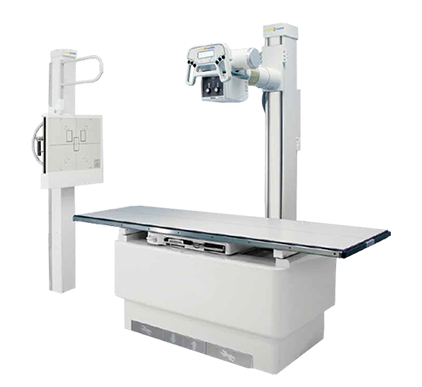
An X-ray machine contains an X-ray tube, which is the source of X-rays. An X-ray tube consists of a cathode and an anode. When a high voltage is applied to the cathode and anode, electrons are accelerated from the cathode to the anode.
X-ray production:
When accelerated electrons strike the anode, X-ray photons are produced. Those x-ray photons have enough energy to travel through the body and interact with a detector or film on the other side. Patient position: The patient is placed between the x-ray tube and the detector or film. The part of the body to be imaged is usually placed in the path of the X-ray beam.
X-ray exposure:
When the X-ray machine is activated, a short beam of X-rays is produced. X-ray photons travel through the body and are attenuated (weakened) in intensity based on the density and composition of the tissues they encounter.
Imaging:
X-ray photons passing through the body reach a detector or film on the other side. The detector converts the X-ray photons into an electrical signal that is processed by a computer to form a digital image. In film-based systems, X-ray photons expose the film, which is then developed to produce a visible image.
Image interpretation:
The resulting X-ray image, called an X-ray, is interpreted by a radiologist or other medical professionals. They will analyze the image to detect any abnormalities, fractures, tumors or other conditions.
It is important to note that X-ray equipment must be used by trained professionals to ensure the safety of both the patient and the operator. Strict protocols and safety measures are followed to minimize radiation and protect the well being of those involved.

